The following document was written by Mr Vik Veer MBBS(lond) MRCS(eng) DoHNS(eng) in Dec 2007. You may use the information here for personal use but if you intend to publish or present it, you must clearly credit the author and www.clinicaljunior.com
This site is not intended to be used by people who are not medically trained. Anyone using this site does so at their own risk and he/she assumes any and all liability. ALWAYS ASK YOUR SENIOR IF YOU ARE UNSURE ABOUT A PROCEDURE. NEVER CONDUCT A PROCEDURE YOU ARE UNSURE ABOUT.
Examination of the Ear
The following document is one way of examining the ear. There are obviously many ways and techniques to do this which aren't mentioned here. I suspect you will only use this as a guide to your own examining technique which you should evolve to suit your own approach and style. I have also assumed that you have some knowledge of ENT throughout this examination. If you are uncertain to the reasons why i have mentioned things in my examination or need clarification there are some excellent books you can refer to. I would suggest using 'Key Topics in Otolaryngology' or 'ENT Secrets', both are adequate for SHO / ST level ENT.
I have written this with the idea that this will be used in an exam setting - so you will be presenting your findings as opposed to a clinic setting. i would try and talk constantly whilst examining the patient. It keeps the examiner interested and shows off what you know.

Ask for consent and then continue with inspection
“Is there any pain at all? (response) and would it be alright if, while i am examining you, that i speak to the examiner about you?"
(remember to wait for response - too often we rehearse these examinations so often that we forget to wait for the answers to these questions in the panic of an exam)
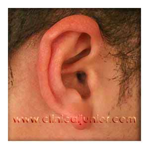
“From infront the ears appear to be of symmetrical shape and position”
Move on to then start with the good ear before going on to the bad ear (explain this is what you normally do to see what is normal for the patient - the examiner may now ask you to move along and examine the pathological ear first.)
“On inspection the post-auricular, mastoid and pre-auricular areas appear normal with no evidence of scars, inflammation, pits or sinuses”
“The pinna appears normal with no obvious scars, inflammation, or deformity”
Ask about pain again!
“Palpation of these areas also appears normal with no tragal, pinna, or mastoid tenderness or masses.”
Pull pinna superiorly, laterally and posteriorly at the same time (in children pull inferiorly and posteriorly)
Comment firstly on the conchal bowl and then the canal itself
“The conchal bowl and external auditory canal appears normal with no evidence of infection or masses”
Now we can comment on the tympanic membrane. Describe what you see in an orderly fashion, going through the various land marks.
“the pars flaccida appears normal with no obvious retraction pocket, keratin masses or any other abnormality. The pars tensa has a normal annulus surrounding it with no marginal perforations or masses.
Occasionally you can't see the whole tympanic membrane, so you can say.....
"I am unable to see the whole tympanic membrane as there is a prominent posterior bulge of the anterior inferior canal wall.
"I can see a normal umbo, and long process of the malleus. The short process of the malleus is visible in the anterior superior quadrant. Also in the posterior superior quadrant I can see the long process of the incus. The anterior and posterior ligaments of the malleus with the corda tympani are also visible and have normal appearances."
"The pars tensa has a typical appearance with no obvious bulging or retraction and therefore has a normal light reflex. The membrane colour and vascularity is also normal.”
“Normally I would refer to the tympanogram to assess the compliance of the tympanic membrane however I could use pneumatic otoscopy to assess the mobility. Otherwise you could use a valsalva for positive pressure and Toynbee’s manoeuvre for negative pressure to assess movement of the tympanic membrane.
Behind the tympanic membrane there aren’t any obvious masses or any collection of fluid or glue.”
If there is pathology then you should describe each quadrant of the tympanic membrane separately
Perforations describe if the perforation is central or marginal (ie involving the annulus) and then which quadrant it is in. You can also assess its size by judging the approximate percentage of the pars tensa (e.g. there is a 20% central posterior-inferior perforation of the pars tensa). If you are able to see into the middle cavity then mention all the structures that you are able to see.
Retraction pockets mention location (pars flaccida or tensa) and whether there is any epithelial debris within it, or if it is a self cleaning pocket.
Typanosclerosis again describe position and extent.
Fistula test
apply tragal pressure (enough to occlude the canal), and whilst pressing and assess the eyes for conjugate deviation of the eyes away from the examined side with the fast component of nystagmus going towards the diseased side.
Hearing tests
Perform free field speech tests using bisyllablic words(spondees) e.g. oatmeal, popcorn, cowboy – or you can use numbers 21, 98, 48 etc. Test with a whispered, conversational and shouted voice (in that order) 60 cm (approximately arms lenght), from the ear.
Tuning fork tests
Use Webers test first by positioning the 512kHz tuning fork firmly on the midline of the forehead and ask where they hear the sound - equally in both ears is normal - a lateralising webers test only really tells you that there is a difference in function between the ears. to get more information you need to move on to Rinne's test.
Rinne's test place the 512kHz tuning fork on one of the mastoid processes firmly and ask if they can hear the sound. When they can, place the tip of the tuning fork 2cm from the external auditory canal and ask, in which position the sound is loudest. Abnormal is when the mastoid (bone conduction) is louder than through the canal (air conduction). It should be remembered that in a patient with very bad hearing on one side, Rinne's test becomes meaningless without masking the opposite side (barany's noise box) as the good ear will pick up the sound on the other side.
Strangely the test is considered "positive" if a normal result is gained (which is the opposite of most medical signs and tests. It is probably safer to say what you found rather than using this confusing system.
“the Weber’s test showed no difference in function between the ears and the Rinne’s test was positive bilaterally indicating air conduction is better than bone conduction.”
You can now attempt to wrap up the examination with some final examination that they may or may not ask you to do.
“I would then normally move on to examination of the cranial nerves particularly the facial nerve and taste sensation. I would complete my examination of the ear by examining the post nasal space in particular the openings of the Eustachian tubes, and examining the neck for any lymphadenopathy.”
"With all these patients i would after my examination ask to see a pure tone audiogram and tympanogram so that i may have a fuller and more objective analysis of this patient's hearing function."